
Prof. Yonghui Zhang’s group reported how Vγ9Vδ2 T cells recognize phosphoantigens
Source:Yonghui Zhang
2019-04-22
Recently, the research group directed by Prof. Yonghui Zhang at Tsinghua University published an article in Immunity, entitled “A structural change in butyrophilin upon phosphoantigen binding underlies phosphoantigen-mediated Vγ9Vδ2 T cell activation”. This study reveals the “inside-out” mechanism of action of phosphoantigens in Vγ9Vδ2 T cell activation at the atomic level, facilitating future immunotherapeutic drug design.
Since the discovery in the mid-1980s, it is now clear that γδ T cells are involved in the control of tissue homeostasis, infection, and malignancy. However, although knowledge of the antigens recognized by αβ T cells has expanded considerably over the past 30 years, little is known about how γδ T cells recognize their ligands. Vγ9Vδ2 T cells, an important set of T cells that are of immunotherapeutic interest, respond to tumors or microbes by recognizing isoprenylated diphosphate metabolites termed as “phosphoantigens”. Vγ9Vδ2 T cells sense phosphoantigens in a manner independent of MHC restrictions. A paper in Nature Immunology in 2013 made a bold claim that the extracellular domain of BTN3A1 was the binding site of phosphoantigens, but this conclusion has not been verified by any other groups working in the area of γδ T cell activation. A highly-cited Immunity paper from Sandstrom et al in 2014 got the field back on track by showing that the intracellular B30.2 domain of butyrophilin 3A1 (BTN3A1) is the binding site for phosphoantigens. To quote Erin Adams in Chicago University, “We have just pulled the curtain back on the inner-workings of Vγ9Vδ2 T cell activation”. However, the understanding of Vγ9Vδ2 T cell activation is far from complete.
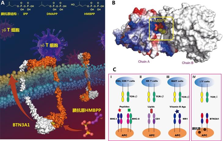
By using a multifaceted approach involving crystallography, molecular dynamics simulations, sedimentations velocity analytical ultracentrifugation (SV-AUC), chemical probes, as well as single-cell force microscopy, Professor Zhang’s group has made an important discovery that deepens understanding of the basic γδ T cell biology. Phosphoantigens bind to the the intracellular B30.2 domain of BTN3A1 in target cells and induces a (β → α) conformational transition of Histidine 351. This conformational change, together with other residue movements induced by HMBPP binding, were conveyed to the juxtamembrane, a process executed by intracellular B30.2 proteins with certain configurations. Phosphoantigen sensing was translated to increased avidity between the extracellular domain of BTN3A1 and the Vγ9Vδ2 TCR, as measured by atomic force microscopy, and led to effective Vγ9Vδ2 T cell activation. These findings provide insight into the molecular underpinnings of Vγ9Vδ2 T cell activation with implications to drug design.
Since the discovery in the mid-1980s, it is now clear that γδ T cells are involved in the control of tissue homeostasis, infection, and malignancy. However, although knowledge of the antigens recognized by αβ T cells has expanded considerably over the past 30 years, little is known about how γδ T cells recognize their ligands. Vγ9Vδ2 T cells, an important set of T cells that are of immunotherapeutic interest, respond to tumors or microbes by recognizing isoprenylated diphosphate metabolites termed as “phosphoantigens”. Vγ9Vδ2 T cells sense phosphoantigens in a manner independent of MHC restrictions. A paper in Nature Immunology in 2013 made a bold claim that the extracellular domain of BTN3A1 was the binding site of phosphoantigens, but this conclusion has not been verified by any other groups working in the area of γδ T cell activation. A highly-cited Immunity paper from Sandstrom et al in 2014 got the field back on track by showing that the intracellular B30.2 domain of butyrophilin 3A1 (BTN3A1) is the binding site for phosphoantigens. To quote Erin Adams in Chicago University, “We have just pulled the curtain back on the inner-workings of Vγ9Vδ2 T cell activation”. However, the understanding of Vγ9Vδ2 T cell activation is far from complete.
By using a multifaceted approach involving crystallography, molecular dynamics simulations, sedimentations velocity analytical ultracentrifugation (SV-AUC), chemical probes, as well as single-cell force microscopy, Professor Zhang’s group has made an important discovery that deepens understanding of the basic γδ T cell biology. Phosphoantigens bind to the the intracellular B30.2 domain of BTN3A1 in target cells and induces a (β → α) conformational transition of Histidine 351. This conformational change, together with other residue movements induced by HMBPP binding, were conveyed to the juxtamembrane, a process executed by intracellular B30.2 proteins with certain configurations. Phosphoantigen sensing was translated to increased avidity between the extracellular domain of BTN3A1 and the Vγ9Vδ2 TCR, as measured by atomic force microscopy, and led to effective Vγ9Vδ2 T cell activation. These findings provide insight into the molecular underpinnings of Vγ9Vδ2 T cell activation with implications to drug design.
