
Post-myocardial infarction immunosuppressive microenvironment driven by lymphatic endothelial transcription factor Tbx1
Source:Zhen Zhang
2023-08-28
The heart is an organ susceptible to autoimmune attacks. These autoimmune attacks play a significant role in various heart diseases such as myocarditis. They may amplify heart injuries through a chain reaction and proceed insidiously to heart failure. In certain cases, they can rapidly expand, leading to a sharp decline in cardiac function and resulting in a higher mortality rate. Epidemiological studies indicate an increased risk of cardiovascular diseases among patients with autoimmune diseases and a worse prognosis following cardiovascular accidents.
Myocardial infarction (MI) poses a serious threat to life and health. Current treatments for myocardial infarction, such as percutaneous coronary intervention, have greatly reduced early mortality after MI. However, MI remains a leading cause of heart failure. Continuous anti-cardiac autoimmune responses following MI play a significant role in the development of post-MI heart failure. Due to a defect in central tolerance, cardiac autoreactive T lymphocytes are continuously released into the periphery. Normally, they are removed through peripheral tolerance. However, under the inflammatory microenvironment induced by ischemic injuries, the peripheral tolerance mediated by dendritic cells is broken. Activated dendritic cells present cardiac-specific antigens released by dead cells, activating autoreactive T lymphocytes specific to these antigens and leading to autoimmunity. Normally, the autoimmunity triggered by MI is transient and will be resolved after the MI-induced inflammation subsides. However, it remains unclear how the MI-induced autoimmunity is restricted.
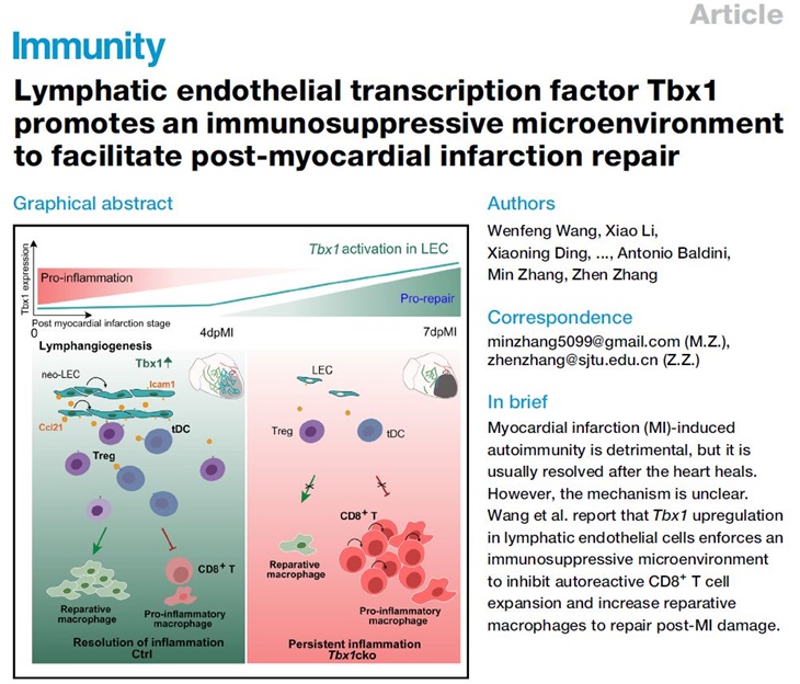
On August 24, 2023, the research group led by Dr. Zhen Zhang from Shanghai Children's Medical Center affiliated with Shanghai Jiao Tong University School of Medicine and their collaborators published a research article titled "Lymphatic endothelial transcription factor Tbx1 promotes an immunosuppressive microenvironment to facilitate post-myocardial infarction repair" in the journal Immunity. This work discovered that the transcription factor Tbx1 is specifically activated in cardiac lymphatic endothelial cells (LECs) after MI. It not only promotes post-MI lymphangiogenesis, facilitating the infiltration of LECs into the infarct area, but also enhances their immunosuppressive function. These two processes effectively establish an immunosuppressive microenvironment in the infarcted area, thereby inhibiting autoreactive CD8+ T cell expansion and promoting the expansion of reparative macrophages to facilitate post-MI repair.
In this study, researchers first identified Tbx1 as one of the most consistently upregulated transcription factors during the period of post-MI 4-7 days. Using the Tbx1LacZ reporter and immunostaining of LEC-specific marker Vegfr3, they revealed that Tbx1 is mainly expressed in cardiac LECs. Tbx1 deficiency in endothelial cells (Tbx1cko) led to impaired neo-lymphangiogenesis in the infarcted area, resulting in reduced LECs in the infarcted zone. Additionally, there was a significant increase in autoreactive CD8+ T cells and a decrease in reparative macrophages. Correspondingly, worsened inflammation and impaired repair were observed in the infarcted area, leading to a significant deterioration in cardiac function.
To understand how LEC Tbx1 regulates autoreactive CD8+ T cells, researchers conducted transcriptomic and epigenetic analyses on enriched post-MI endothelial cells. They found that Tbx1 regulates two categories of gene expression: one that promotes lymphangiogenesis and another with immunomodulatory functions, which includes the cytokine Ccl21 and the surface adhesion molecule Icam1. The Ccl21 receptor Ccr7 is specifically expressed in immune-tolerant dendritic cells (tDC), while the Icam1 receptor Itgal is highly expressed in regulatory CD4+ T cells (Treg) that promote immune tolerance. Multiplex RNA in situ hybridization demonstrated significant spatial co-localization of LECs, tDCs, and CD8+ T lymphocytes in the infarcted area. Tbx1 deficiency resulted in an increased distance between these cell types and LECs. This suggests a pivotal role of cardiac LECs in establishing an immunosuppressive microenvironment in the infarcted area.
To further validate the immunosuppressive roles of Ccl21 and Icam1 mediated by LECs, researchers used Ccl21 blocking antibodies and LEC-specific knockout of Icam1. They found that the absence of Ccl21 or Icam1 significantly reduced the numbers of tDCs or Tregs in hearts, respectively, while both resulted in a significant increase in CD8+ T cell numbers and a notable decrease in cardiac function. Conversely, injecting CD28SA antibodies to expand Tregs in Tbx1cko mice reduced the number of CD8+ T cells and rescued the decreased cardiac function. These results demonstrate that the immunosuppressive effect of post-MI LECs is mediated by Tbx1-driven upregulation of Ccl21 and Icam1 expression.
In summary, this work reveals a mechanism of active suppression of post-MI autoimmunity. LECs play a crucial role in inhibiting autoreactive CD8+ T cells. The discovery of this molecular mechanism provides a novel approach for treating MI and preventing post-MI heart failure. TBX1 happloinsufficiency is a major contributor to congenital heart diseases seen in 22q11.2 deletion syndrome (22q11.2DS), the most common chromosomal microdeletion syndrome with an incidence of up to 1 in 2000 live birth. With advancements in pediatric care, the population of adult 22q11.2DS patients is steadily increasing. Our research indicates that the MI prognosis in this specific population might be worse, which demands preventive treatment for these patients.
Drs. Zhen Zhang and Min Zhang from Shanghai Children's Medical Center affiliated with Shanghai Jiao Tong University School of Medicine are the co-corresponding authors of this research article. The co-first authors include Dr. Wenfeng Wang from Shanghai Jiao Tong University School of Medicine, Dr. Xiao Li from the Texas Heart Institute, and Dr. Xiaoning Ding and Shanshan Xiong from Shanghai Jiao Tong University School of Medicine. Dr. Chuanxin Huang from the Shanghai Institute of Immunology provided a great deal of guidance and assistance in this research.
Article Links: https://www.sciencedirect.com/science/article/pii/S1074761323003321
Myocardial infarction (MI) poses a serious threat to life and health. Current treatments for myocardial infarction, such as percutaneous coronary intervention, have greatly reduced early mortality after MI. However, MI remains a leading cause of heart failure. Continuous anti-cardiac autoimmune responses following MI play a significant role in the development of post-MI heart failure. Due to a defect in central tolerance, cardiac autoreactive T lymphocytes are continuously released into the periphery. Normally, they are removed through peripheral tolerance. However, under the inflammatory microenvironment induced by ischemic injuries, the peripheral tolerance mediated by dendritic cells is broken. Activated dendritic cells present cardiac-specific antigens released by dead cells, activating autoreactive T lymphocytes specific to these antigens and leading to autoimmunity. Normally, the autoimmunity triggered by MI is transient and will be resolved after the MI-induced inflammation subsides. However, it remains unclear how the MI-induced autoimmunity is restricted.
On August 24, 2023, the research group led by Dr. Zhen Zhang from Shanghai Children's Medical Center affiliated with Shanghai Jiao Tong University School of Medicine and their collaborators published a research article titled "Lymphatic endothelial transcription factor Tbx1 promotes an immunosuppressive microenvironment to facilitate post-myocardial infarction repair" in the journal Immunity. This work discovered that the transcription factor Tbx1 is specifically activated in cardiac lymphatic endothelial cells (LECs) after MI. It not only promotes post-MI lymphangiogenesis, facilitating the infiltration of LECs into the infarct area, but also enhances their immunosuppressive function. These two processes effectively establish an immunosuppressive microenvironment in the infarcted area, thereby inhibiting autoreactive CD8+ T cell expansion and promoting the expansion of reparative macrophages to facilitate post-MI repair.
In this study, researchers first identified Tbx1 as one of the most consistently upregulated transcription factors during the period of post-MI 4-7 days. Using the Tbx1LacZ reporter and immunostaining of LEC-specific marker Vegfr3, they revealed that Tbx1 is mainly expressed in cardiac LECs. Tbx1 deficiency in endothelial cells (Tbx1cko) led to impaired neo-lymphangiogenesis in the infarcted area, resulting in reduced LECs in the infarcted zone. Additionally, there was a significant increase in autoreactive CD8+ T cells and a decrease in reparative macrophages. Correspondingly, worsened inflammation and impaired repair were observed in the infarcted area, leading to a significant deterioration in cardiac function.
To understand how LEC Tbx1 regulates autoreactive CD8+ T cells, researchers conducted transcriptomic and epigenetic analyses on enriched post-MI endothelial cells. They found that Tbx1 regulates two categories of gene expression: one that promotes lymphangiogenesis and another with immunomodulatory functions, which includes the cytokine Ccl21 and the surface adhesion molecule Icam1. The Ccl21 receptor Ccr7 is specifically expressed in immune-tolerant dendritic cells (tDC), while the Icam1 receptor Itgal is highly expressed in regulatory CD4+ T cells (Treg) that promote immune tolerance. Multiplex RNA in situ hybridization demonstrated significant spatial co-localization of LECs, tDCs, and CD8+ T lymphocytes in the infarcted area. Tbx1 deficiency resulted in an increased distance between these cell types and LECs. This suggests a pivotal role of cardiac LECs in establishing an immunosuppressive microenvironment in the infarcted area.
To further validate the immunosuppressive roles of Ccl21 and Icam1 mediated by LECs, researchers used Ccl21 blocking antibodies and LEC-specific knockout of Icam1. They found that the absence of Ccl21 or Icam1 significantly reduced the numbers of tDCs or Tregs in hearts, respectively, while both resulted in a significant increase in CD8+ T cell numbers and a notable decrease in cardiac function. Conversely, injecting CD28SA antibodies to expand Tregs in Tbx1cko mice reduced the number of CD8+ T cells and rescued the decreased cardiac function. These results demonstrate that the immunosuppressive effect of post-MI LECs is mediated by Tbx1-driven upregulation of Ccl21 and Icam1 expression.
In summary, this work reveals a mechanism of active suppression of post-MI autoimmunity. LECs play a crucial role in inhibiting autoreactive CD8+ T cells. The discovery of this molecular mechanism provides a novel approach for treating MI and preventing post-MI heart failure. TBX1 happloinsufficiency is a major contributor to congenital heart diseases seen in 22q11.2 deletion syndrome (22q11.2DS), the most common chromosomal microdeletion syndrome with an incidence of up to 1 in 2000 live birth. With advancements in pediatric care, the population of adult 22q11.2DS patients is steadily increasing. Our research indicates that the MI prognosis in this specific population might be worse, which demands preventive treatment for these patients.
Drs. Zhen Zhang and Min Zhang from Shanghai Children's Medical Center affiliated with Shanghai Jiao Tong University School of Medicine are the co-corresponding authors of this research article. The co-first authors include Dr. Wenfeng Wang from Shanghai Jiao Tong University School of Medicine, Dr. Xiao Li from the Texas Heart Institute, and Dr. Xiaoning Ding and Shanshan Xiong from Shanghai Jiao Tong University School of Medicine. Dr. Chuanxin Huang from the Shanghai Institute of Immunology provided a great deal of guidance and assistance in this research.
