


Qiang Liu's Team Uncovers a Novel Mechanism by Which Meningeal ILC2s Drive Neuroinflammation, Offering a New Therapeutic Target for Multiple Sclerosis
Source:Qiang Liu
2026-10-22
On August 6, 2026, the research team led by Professor Liu Qiang at Tianjin Medical University General Hospital, in collaboration with the team led by Professor Zhou Jie, published a research article online in the Cell Press journal Immunity, entitled "Group 2 innate lymphoid cells activate myelin-reactive T cells to augment neuroinflammation". The study reveals that group 2 innate lymphoid cells (ILC2s) accumulate in the meninges during neuroinflammation, where they activate and expand myelin-reactive T cells through MHC II-mediated antigen presentation, driving central nervous system (CNS) inflammation and demyelination. These findings suggest that targeting meningeal ILC2s could offer a promising new strategy for the treatment of autoimmune diseases of the nervous system.
Multiple sclerosis (MS) is an autoimmune disease of the CNS characterized pathologically by myelin damage, axonal degeneration, and persistent inflammation. Most patients experience their first onset between the ages of 20 and 40, making MS one of the leading causes of disability among young and middle-aged adults and imposing a heavy economic and psychological burden. Recent studies have highlighted the increasingly prominent role of the meninges, the membranes that envelop the brain and spinal cord, in CNS immunity: T cells capable of attacking myelin are first activated in the meningeal region and then enter the brain tissue, where they trigger spreading inflammation and demyelinating injury. However, which specific immune cells in the meninges drive the activation of myelin-reactive T cells has remained unclear.
Innate lymphoid cells (ILCs) are a newly recognized class of immune cells that do not depend on antigen-specific recognition and can rapidly sense the tissue microenvironment. Among them, ILC2s reside mainly in mucosal barrier tissues such as the intestine, lung, and skin, where they regulate allergic responses, tissue repair, and defense against infection. In previous collaborative work on acute CNS injury, the team found that ILC2s resident in the brain can shape intracerebral inflammation and neural repair after brain injury by producing molecules such as amphiregulin (Areg) (Neuron, 2025). However, whether ILC2s influence the activation of myelin-reactive T cells in autoimmune neuroinflammation has remained an open question.
Using the experimental autoimmune encephalomyelitis (EAE) model, single-cell sequencing and flow cytometry analyses showed that ILC2s accumulate markedly in the dura mater and display a distinctive transcriptional profile: MHC II and antigen-processing-related genes are significantly upregulated, whereas canonical type 2 immunity signature genes are downregulated. Tissue immunofluorescence staining revealed that ILC2s are located in close proximity to CD4+ T cells in the dura mater, suggesting direct interactions between the two cell types. Genetic deficiency or specific depletion of ILC2s significantly alleviated neurological deficits and demyelination in EAE mice and reduced the accumulation of endogenous pathogenic T cells in the CNS. Single-cell TCR sequencing showed that the absence of ILC2s markedly restricted the clonal expansion of myelin-reactive T cells in the dura mater. Mechanistically, ILC2s can induce the proliferation of myelin-specific T cells through MHC II; conditional knockout of MHC II in ILC2s significantly attenuated T cell activation and neuroinflammation, confirming that ILC2s act as "antigen-presenting cells" to drive the expansion of myelin-reactive T cells. Notably, the incidence of MS is significantly higher in women than in men. The researchers found that female EAE mice harbor significantly more dural ILC2s with higher MHC II expression than males, and that the sex difference in disease was abolished after MHC II was knocked out in ILC2s. Clinical samples corroborated these findings: MHC II expression on peripheral blood ILC2s was significantly elevated in patients with MS and was closely correlated with MS disease activity.
This study uncovers a pathogenic role for meningeal ILC2s in autoimmune neuroinflammation and broadens the theoretical understanding of CNS border immunity, particularly meningeal immunity. It reveals that ILC2s are not merely "guardians" of tissue repair: under specific conditions they can switch into "accelerators" of neuroinflammation, amplifying pathogenic T cell responses through antigen presentation. These findings provide an entirely new perspective for understanding the pathogenesis of MS and its sex differences. Targeting meningeal ILC2s and their antigen-presenting function holds promise as a new strategy for limiting CNS inflammation and autoimmunity.
Professor Liu Qiang's team has long been dedicated to research on the immune mechanisms of brain diseases and their clinical intervention. The team has led multiple clinical studies of immune-based interventions for neurological disorders, developed a fully human BAFFR monoclonal antibody and CAR-T cells, and launched investigator-initiated trials, demonstrating broad prospects for translational application.
Article link: https://www.cell.com/immunity/abstract/S1074-7613(26)00310-9
Multiple sclerosis (MS) is an autoimmune disease of the CNS characterized pathologically by myelin damage, axonal degeneration, and persistent inflammation. Most patients experience their first onset between the ages of 20 and 40, making MS one of the leading causes of disability among young and middle-aged adults and imposing a heavy economic and psychological burden. Recent studies have highlighted the increasingly prominent role of the meninges, the membranes that envelop the brain and spinal cord, in CNS immunity: T cells capable of attacking myelin are first activated in the meningeal region and then enter the brain tissue, where they trigger spreading inflammation and demyelinating injury. However, which specific immune cells in the meninges drive the activation of myelin-reactive T cells has remained unclear.
Innate lymphoid cells (ILCs) are a newly recognized class of immune cells that do not depend on antigen-specific recognition and can rapidly sense the tissue microenvironment. Among them, ILC2s reside mainly in mucosal barrier tissues such as the intestine, lung, and skin, where they regulate allergic responses, tissue repair, and defense against infection. In previous collaborative work on acute CNS injury, the team found that ILC2s resident in the brain can shape intracerebral inflammation and neural repair after brain injury by producing molecules such as amphiregulin (Areg) (Neuron, 2025). However, whether ILC2s influence the activation of myelin-reactive T cells in autoimmune neuroinflammation has remained an open question.
Using the experimental autoimmune encephalomyelitis (EAE) model, single-cell sequencing and flow cytometry analyses showed that ILC2s accumulate markedly in the dura mater and display a distinctive transcriptional profile: MHC II and antigen-processing-related genes are significantly upregulated, whereas canonical type 2 immunity signature genes are downregulated. Tissue immunofluorescence staining revealed that ILC2s are located in close proximity to CD4+ T cells in the dura mater, suggesting direct interactions between the two cell types. Genetic deficiency or specific depletion of ILC2s significantly alleviated neurological deficits and demyelination in EAE mice and reduced the accumulation of endogenous pathogenic T cells in the CNS. Single-cell TCR sequencing showed that the absence of ILC2s markedly restricted the clonal expansion of myelin-reactive T cells in the dura mater. Mechanistically, ILC2s can induce the proliferation of myelin-specific T cells through MHC II; conditional knockout of MHC II in ILC2s significantly attenuated T cell activation and neuroinflammation, confirming that ILC2s act as "antigen-presenting cells" to drive the expansion of myelin-reactive T cells. Notably, the incidence of MS is significantly higher in women than in men. The researchers found that female EAE mice harbor significantly more dural ILC2s with higher MHC II expression than males, and that the sex difference in disease was abolished after MHC II was knocked out in ILC2s. Clinical samples corroborated these findings: MHC II expression on peripheral blood ILC2s was significantly elevated in patients with MS and was closely correlated with MS disease activity.
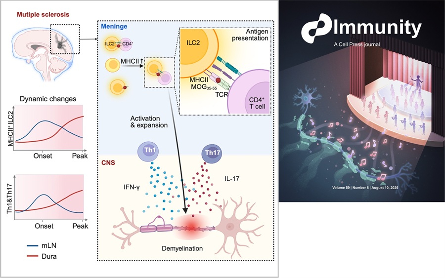
Figure. ILC2s drive neuroinflammation by activating myelin-reactive T cells.
Left panel: ILC2s accumulate in the meningeal region, where they activate and promote the expansion of myelin-reactive T cells, driving CNS inflammation and demyelinating injury.
Right panel: Candidate cover image for Immunity. ILC2s shape the progression of CNS inflammation and demyelination by finely tuning T cell responses.
This study uncovers a pathogenic role for meningeal ILC2s in autoimmune neuroinflammation and broadens the theoretical understanding of CNS border immunity, particularly meningeal immunity. It reveals that ILC2s are not merely "guardians" of tissue repair: under specific conditions they can switch into "accelerators" of neuroinflammation, amplifying pathogenic T cell responses through antigen presentation. These findings provide an entirely new perspective for understanding the pathogenesis of MS and its sex differences. Targeting meningeal ILC2s and their antigen-presenting function holds promise as a new strategy for limiting CNS inflammation and autoimmunity.
Professor Liu Qiang's team has long been dedicated to research on the immune mechanisms of brain diseases and their clinical intervention. The team has led multiple clinical studies of immune-based interventions for neurological disorders, developed a fully human BAFFR monoclonal antibody and CAR-T cells, and launched investigator-initiated trials, demonstrating broad prospects for translational application.
Article link: https://www.cell.com/immunity/abstract/S1074-7613(26)00310-9
